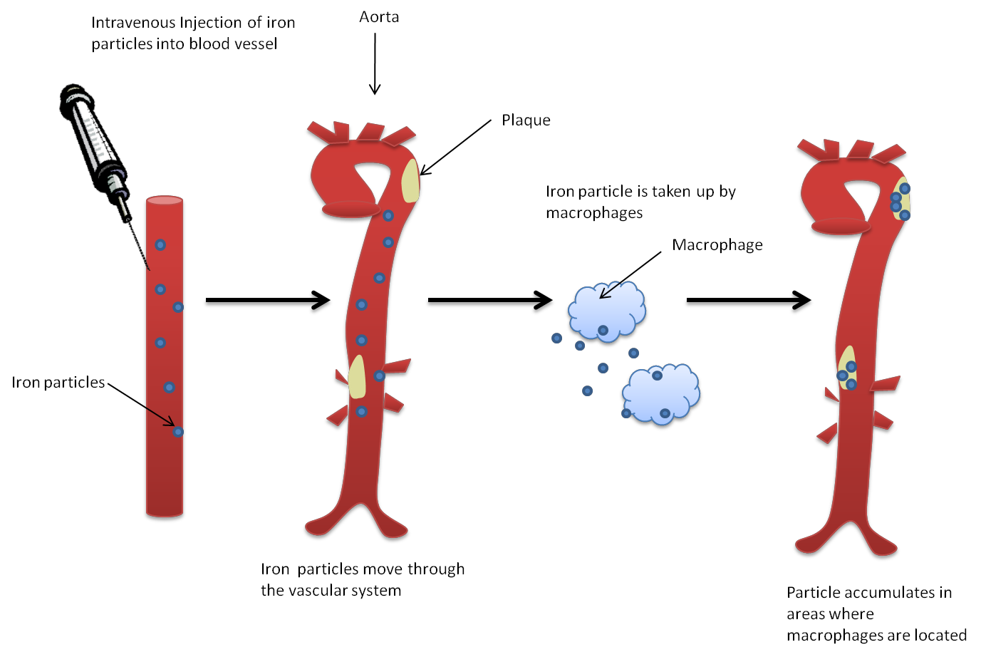
Radiation therapy can accelerate atherosclerotic changes. Macrophages and monocytes play an important role in atherosclerosis and plaque behaviors. Inflammation within these plaques can be quantitatively assessed by measuring T2* value of these plaques following intravenous administration of super paramagnetic iron oxide nanoparticles. From the cohort of pancreatic cancer patient who underwent MRI scans before and after administration of super paramagnetic iron oxide nanoparticles, we will identify two groups A)patients who underwent neoadjuvant therapy prior to undergoing these MRI scans and B)patients who did not receive neoadjuvant therapy. Quantitative T2* measurements of aortic wall and atherosclerotic plaques will be performed and compared in each group.