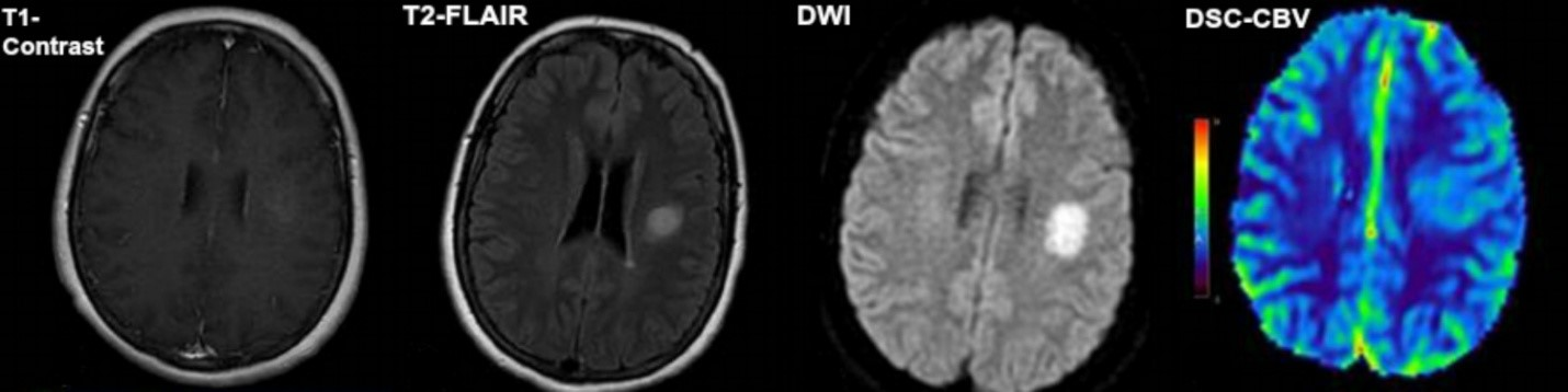
My project will focus on assessing MRI measurement reproducibility in patients with oligodendroglioma. Oligodendrogliomas derive from oligodendrocytes, a glial cell that protects neurons in the central nervous system. Since oligodendroglioma is a relatively slow-growing cancer, neuro-oncologists refrain from surgery in favor of a “watchful waiting” tactic based on imaging results. Brain tumors are measured according to the Response Assessment in Neuro-Oncology (RANO) criteria. Radiologists measure the product of the two longest perpendicular diameters of the tumor visible in the post-contrast T1-weighted MR slice in which the tumor is found to be of the largest size. Tumor size is monitored from the start of treatment through each subsequent imaging session, and is then classified into one of four response categories based on relative size change: complete response, partial response, stable disease, or progressive disease. However, there is significant variability in patients’ tumor size measurements, arising from intra- and inter-observer variability and scanner differences. The aim of this project is to define the observer and scanner variability in quantitative tumor biomarkers based on perfusion, diffusion, and anatomical MRI. Multiple readers will analyze axial MR slices from oligodendroglioma patients with each of the following parameters: post-contrast T1-weighted, FLAIR T2-weighted, DWI-ADC, and DSC-CBV. The readers will measure the lesions in the same images at two different time points to assess intra-observer variability. We hypothesize that intra-/inter-observer and intra-/inter-scanner variability will not affect assessment of the oligodendroglioma progression as defined by RANO. This project will establish baseline variances in tumor measurements so that researchers can better classify tumor volume change as either genuine tumor response/progression or simply measurement variance.