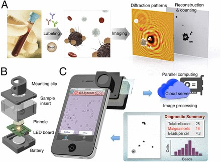
Much of the developing world currently has limited availability of pathology services resulting in numerous cases of undetected and untreated diseases that, if caught earlier on in their progression, would be much easier and less expensive to treat. A group of researchers at the Center for Systems Biology at Massachusetts General Hospital has developed a device called the D3 (digital diffraction diagnostic) system that can be used to diagnose various physiological conditions including lymphoma, cervical cancer, and HPV in point-of-care settings using a smartphone. This device, which attaches itself to the lens of a smartphone, consists of a battery-powered LED light, circuit board, sample insert, pinhole, and mounting clip and utilizes the smartphone camera to take an image of a manually-inserted blood or tissue sample. Before the image is taken, the sample is labeled using microbeads coated with ligands (e.g. antibodies or nucleotides) that bind to specific biomarkers indicative of cancer or HPV. In order to detect lymphoma, cells from a fine-needle aspirate (FNA) of a lymph node are captured by primary antibody CD20 coated on a glass coverslip, then microbeads coated with either kappa or lambda antibody are added to bind to the cells and interrogate their polarization. A similar process is carried out for diagnosing HPV. However, the viral DNA is targeted by microbeads rather than the cancer cells containing the CD20 antigen. Once the image is taken, it is uploaded via a secure, encrypted cloud network to a database at MGH where the image is analyzed using an algorithm that detects the presence of cancer or HPV by identifying the unique diffraction patterns of the bound beads as well as their proximity to the analyte of interest. Based on the number of microbeads bound to target cells, a patient can be quickly, inexpensively, and accurately diagnosed. The D3 device has detected the presence of tumor proteins in cancer cell lines as accurately as the gold standard of microscopy and flow cytometry and allows for an analysis of up to 100,000 cells at a time. The goal of this project is to refine and optimize the D3 assay in order to create an efficient diagnostic device capable of revolutionizing pathology services related to cancer and other diseases in developing countries.