Characterization of Targeted PARP-inhibitor Nanoformulations In Vitro and In Vivo
Mentor: Srinivas Sridhar, Ph.D.
Institution: Northeastern University
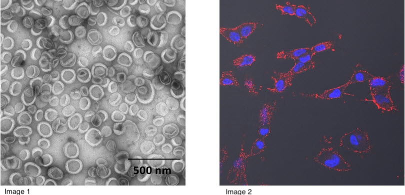
[Image 1] NanoTLZ stained with 1.5% uranyl acetate as viewed by transmission electron microscopy. [Image 2] Visualization of nanoparticle uptake in vitro via confocal microscopy. MDA-MB-231 cells were incubated with Cy5 tagged NanoTLZ (red) to visualize nanoparticle uptake in vitro via confocal microscopy. Nucleus staining was done with Hoechst 33342 dye (blue).